2024: The Exercise Gap Medicine Cannot Fill
Picture a typical orthopedic waiting room.
A woman in her seventies who avoids stairs because her knees ache. A man in his sixties whose fear of falling makes any vigorous movement feel dangerous. A middle-aged patient whose cardiovascular condition has ruled out strenuous exercise entirely. The physician’s advice is invariably the same:
“You need to exercise. Resistance training, especially.”
This is the difficult reality of sarcopenia exercise: the people who need resistance training most are often the ones who struggle the most to perform it consistently.
For patients with joint pain, diminished cardiopulmonary function, or no access to supervised training, the actual adherence rate for resistance exercise programs is extremely low. The prescription exists — the ability to follow it often does not. This gap is the defining clinical challenge of sarcopenia management today.
According to the 2024 Korea National Health and Nutrition Examination Survey, nearly one in ten Koreans aged 65 and older — 9.4% — already has sarcopenia. The prevalence is rising every year. Sarcopenia is not simply a feature of aging. Since 2016, it has carried its own ICD-10 diagnostic code — M62.84 — a recognized clinical disease that directly drives falls, fractures, metabolic dysfunction, and dependency on long-term care.
This is where sarcopenia exercise support becomes important. The technology designed to help close this gap is the WME-01 Wave Motion Core — a PEMF + Sonic Dual Wave full-body exercise device.
Sarcopenia Exercise Support: Two Waves, Two Pathways
What distinguishes the Wave Motion Core from conventional exercise equipment is straightforward: a single device incorporates two technologies, each stimulating a different physiological pathway simultaneously.
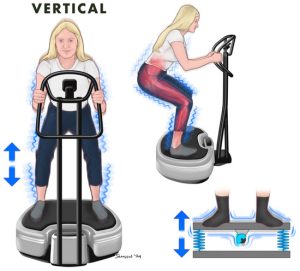
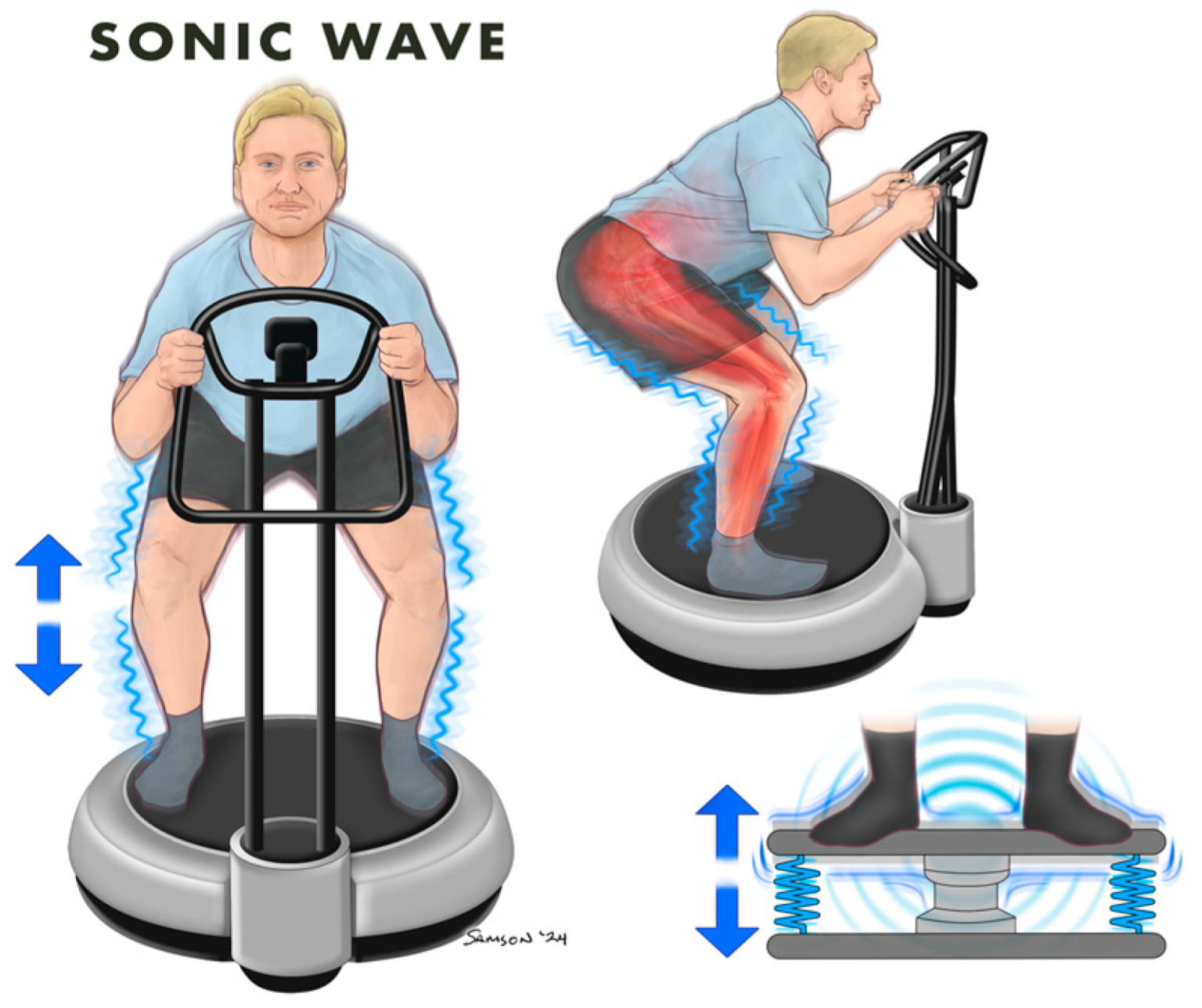
Sonic Wave delivers acoustic vibration at audible frequencies directly into body tissue. Unlike conventional whole-body vibration (WBV) devices — which use mechanical motors to physically oscillate a platform up and down — sonic wave technology uses a transducer to transfer acoustic energy directly into tissue. This enables neuromuscular stimulation without joint impact loading.
PEMF (Pulsed Electromagnetic Field) operates at an entirely different level. While sonic wave acts on the nervous and muscular system, PEMF penetrates tissue and acts directly at the cellular level — on cell membranes and mitochondria.
The two mechanisms are independent. They work simultaneously.
Mechanism 1 | Sonic Wave: The Science of Vibration-Activated Neuromuscular Stimulation
The clinical research world has spent decades establishing how vibration stimulation affects muscle tissue — primarily through studies using mechanical whole-body vibration (WBV) platforms. The fundamental principles these studies revealed apply regardless of how vibration is delivered.
When vibratory stimulation reaches the body, afferent sensory receptors and muscle spindles in the sole of the foot and lower limb detect it. This signal travels up the spinal cord and is transmitted to the anti-gravity muscles of the lower extremity — the quadriceps, gastrocnemius, and others — inducing involuntary recruitment of muscle fibers. This is the neurophysiological basis for muscular response without active volitional effort and without joint loading.
What has this principle produced in actual clinical settings?
📚 Beom J, Lim JY, Lee SY. Sci Rep. 2026. doi:10.1038/s41598-026-45710-y → Systematic review and meta-analysis of 6 RCTs (n=202) in older adults with sarcopenia. Pooled SMD 0.50 (95% CI 0.21–0.80, p=0.001). Significant lower-limb muscle strength improvement vs. control. I²=0%. 🔗 Nature full text
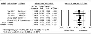
Beom et al. (2026) — Forest Plot, lower-limb muscle strength SMD visualization
📚 Zhuang M, et al. Sci Rep. 2025;15(1):6981 → 12-week RCT (3×/week, n=27) in sarcopenic adults aged 65+. Vibration training group achieved comparable gains in knee extensor strength and gait speed to resistance training group, with superior adherence and safety profiles. 🔗 PubMed
📚 Zafar T, et al. J Clin Med. 2024;13(6):1639 → Meta-analysis of high-quality RCTs (PEDro ≥5). Vibration exercise produced significant improvements across all measured outcomes in chronic low back pain patients: pain, balance, and quality of life. 🔗 PMC
⚠️ Editorial Note: The three studies above were conducted using mechanical WBV platforms. It is scientifically reasonable to expect that WME-01’s sonic wave mechanism activates the same neurophysiological pathways to produce similar outcomes; however, the evidence base will strengthen further as clinical research using sonic wave devices specifically accumulates.
Frequency Is the Critical Variable — Why WME-01’s 5–30Hz Range Aligns With Clinical Guidance
Not all vibration stimulation is equally effective. The question of which frequency range produces the best outcomes in older adults was addressed by a large-scale 2025 network meta-analysis.
📚 Arch Phys Med Rehabil. 2025 — Network meta-analysis (27 RCTs, n=1,608, mean age 73.3 years)
This study directly compared three frequency bands in older adult populations. The findings:
| Outcome Domain | Optimal Frequency Range |
| Static balance | Medium frequency (MF, 20–30Hz) — SUCRA 98.5% |
| Dynamic balance & gait | High frequency (HF) advantage |
| Muscle strength | Superior when combined with traditional resistance training |
WME-01 operates across a 5–30Hz range — encompassing the medium-frequency band identified as most effective for static balance improvement. The ability to adjust across the full 5–30Hz spectrum, rather than being locked to a single frequency, enhances clinical flexibility across different user profiles and therapeutic goals.
Mechanism 2 | PEMF: Activating Ca²⁺ at the Cellular Level
Where sonic wave operates in the neuromuscular system, PEMF acts independently at a deeper level — directly on the cell membrane and mitochondria.
When a pulsed electromagnetic field reaches the cell membrane, the following cascade is initiated:
① Voltage-gated Ca²⁺ channel activation → ② Intracellular Ca²⁺ concentration increases → ③ Calmodulin (CaM) binding → ④ Nitric oxide (NO) production → ⑤ cGMP pathway activation → ⑥ Muscle cell regeneration · microcirculatory expansion · mitochondrial biogenesis
📚 Mittermayr R, et al. — PEMF cellular mechanism (Ca²⁺ → NO → cGMP full cascade) schematic 🔗 Full text
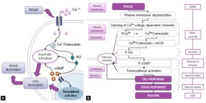
Mittermayr et al. — Ca²⁺ influx through complete NO → cGMP cascade arrow diagram
Clinical Evidence for Sarcopenia Exercise Support Using PEMF
This pathway does not remain at the level of cellular hypothesis. Direct clinical evidence exists.
At the Cellular Level: Regenerative Proteins Are Actually Expressed
📚 Siwak M, et al. Int J Mol Sci. 2023;24(24):17399 → Following 1.5mT PEMF stimulation, skeletal muscle cells (SkMC) demonstrated significant increases in proliferation rate and confirmed expression of muscle regeneration proteins Pax7 and MyoD. 🔗 PMC
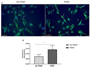
Siwak et al. (2023) — Fluorescence microscopy comparison of SkMC MyoD expression, NO PEMF vs PEMF conditions
In Sarcopenia Patients: +42% Knee Strength, 19% TUG Improvement
📚 Sardinha Leonardo P, et al. (including Lopes-Martins RAB). Life. 2025;15(7):1111. PMC12300015 → 12-session PEMF intervention in older adults with sarcopenia. Results:
- Knee extensor strength: 13.05 → 18.56 kgf (+42%, p<0.001)
- Timed Up and Go (TUG) test: 23.1 → 18.7 seconds (19% reduction, p=0.048)
- SARC-F + CC score: 11.6 → 6.5 (p<0.001) 🔗 PMC
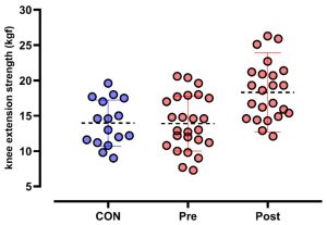
Sardinha Leonardo et al. (2025) — Knee extensor strength dot plot, CON vs Pre vs Post
This is the clinical significance of PEMF. A signal that begins at the cell membrane translates — through multiple simultaneous pathways — into measurable gains in muscle strength.
Two Mechanisms in One Device — The Clinical Meaning of Dual-Wave Technology
| Sonic Wave | PEMF | |
| Site of action | Neuromuscular system (tissue level) | Cell membrane · mitochondria (cellular level) |
| Primary effects | Muscle spindle stimulation → fiber recruitment / microcirculation | Ca²⁺ activation → NO production → muscle cell regeneration |
| vs. conventional | Vibration without impact or noise | Cellular penetration without surface contact |
| Clinical evidence | Vibration meta-analyses (strength, balance, falls) | PEMF sarcopenia RCT (strength, TUG — significant improvement) |
These two mechanisms operate independently across different biological pathways, converging on the same outcome: muscle cells that survive, contract, and regenerate.
Conclusion: The Most Realistic Answer to the Exercise Gap
The central challenge of sarcopenia exercise can be distilled to a single requirement: an intervention that is effective, safe, and sustainable.
The Wave Motion Core activates two independent physiological pathways simultaneously — from the cellular level (Ca²⁺ activation → NO production → muscle cell regeneration) to the tissue level (muscle spindle stimulation → lower-limb fiber recruitment). For patients whose knees ache, whose cardiopulmonary reserve is limited, or for whom active exercise is simply not feasible.
Ten minutes a day. Standing. Or seated.
Muscle continues to age with every passing year. The clinical evidence for intervening in that process already exists.
👉 Explore the Hue Light Wave Motion WME-01 for Sarcopenia Exercise Support
Disclaimer: This device is classified as exercise equipment (general consumer product), not a medical device. Individuals with pre-existing medical conditions should consult a qualified healthcare professional before use.
References
- Korea Disease Control and Prevention Agency. 2024 Korea National Health and Nutrition Examination Survey. 2025.
- Beom J, Lim JY, Lee SY. Sci Rep. 2026. doi:10.1038/s41598-026-45710-y
- Zhuang M, et al. Sci Rep. 2025;15(1):6981. PMID: 40011687
- Zafar T, et al. J Clin Med. 2024;13(6):1639. PMID: 38541865
- Network meta-analysis (27 RCTs, n=1,608). Arch Phys Med Rehabil. 2025.
- Mittermayr R, et al. Mechanisms of PEMF in Medicine. ResPubl Journals.
- Siwak M, et al. Int J Mol Sci. 2023;24(24):17399. PMC10706358
- Sardinha Leonardo P, et al. Life. 2025;15(7):1111. PMC12300015